
Science popularization
Our eye structure is mainly divided into three parts: eyeball, accessory organ of eyeball, optic nerve and blood vessel.
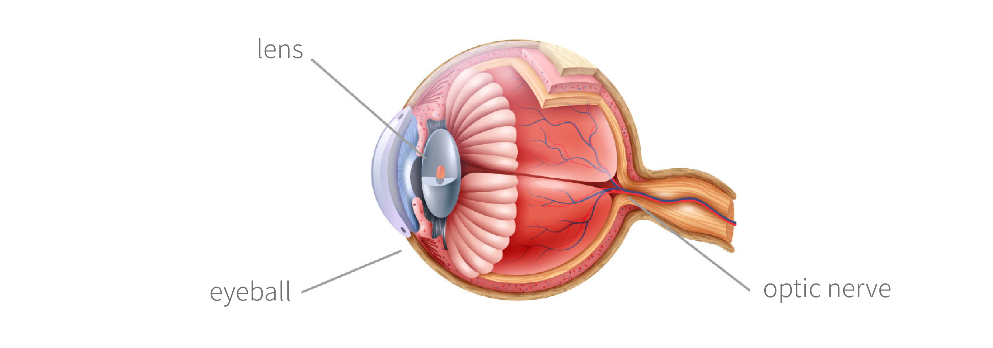
The lens
The transparent lens located behind the pupil and suspended by the suspensory ligament behind the iris in front of the vitreous. The shape of the lens, controlled by the ciliary muscle, changes its thickness and adjusts the refraction of light, focusing light on the retina.
What is cataract
What is cataract
Cataract is a kind of lens in the structure of the eyeball. It is a process of gradual atomization and hardening. The lens changes from a clear and transparent state to a turbid one, which may start from the middle or the side. The lens is located between the iris and the vitreous. The normal lens is in a clear and transparent state. It can adjust the light, focus the light on the retina, and make the eye see a clear image. When the lens gradually becomes turbid and presents brown or white, the penetration of the light will be affected, and the retina will be hindered from receiving a clear image, resulting in visual blur.
Common cataract surgery
Intracapsular cataract extraction: the sclera edge was cut open with a large incision, and after entering the sclera, the crystal was frozen with a frozen head, and the crystal was removed by pulling outward, causing rupture of the suspensory ligament.
Extracapsular cataract extraction: the incision was smaller than the intracapsular extraction, the cloudy nucleus was expelled, the cortex was sucked out, but the posterior capsular was left. The posterior capsule is retained and the posterior chamber intraocular lens can be implanted at the same time. Visual function can be restored immediately after surgery. Therefore, extracapsular cataract extraction has become a routine cataract operation.
Cataract phacoemulsification: it is a new cataract operation in recent years. The nucleus of the lens was shattered by ultrasound to make it chyloid, and then aspirated together with the cortex. The posterior capsule of the lens was retained after surgery, and the atrial iOL could be implanted simultaneously. Its advantages are small incision, less tissue damage, short operation time, quick visual recovery.
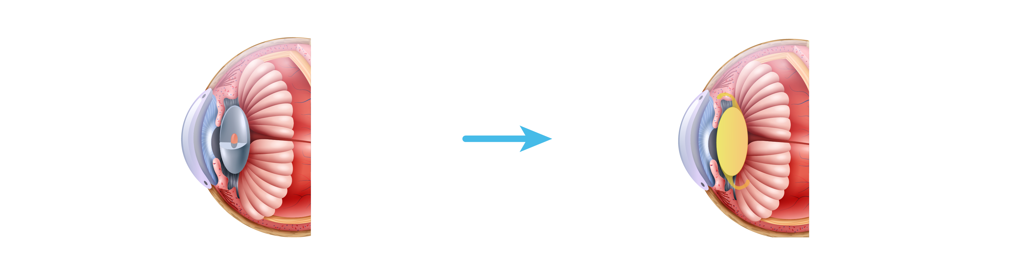
What is intraocular lens
Intraocular lens (IOL)
Intraocular lens is special lens made of synthetic materials.
Types of intraocular lens
According to the different functions of the intraocular lens, it can be divided into the following categories:
- Single focus intraocular lens:After implantation, patients need to wear presbyopia glasses or corrective glasses to obtain clear near or long distance vision.
- aspherical intraocular lens :The main purpose is to reduce the degree of aberrations, improve the visual image quality, and increase the safety of activities in dark light or at night.
- Yellow intraocular lens:To reduce the damage of harmful blue light to retina.
- Correctable astigmatic intraocular lens:It is used to correct the existing corneal astigmatism, effectively reduce astigmatism, and make the postoperative vision of patients clearer.
- Multifocal intraocular lens:Using a circle by circle design, we can make the distance of seeing far, middle and near have different focal length, which can provide better eyesight of presbyopia and near eyesight at the same time, but it will reduce the quality of retinal imaging, and under the condition of the case light, there will be the feeling of glare and halo, which may cause inconvenience to the patients who often need to drive at night.
- Adjustable intraocular lens:Designed according to the principle of human eye regulation, it can correct cataract and presbyopia at the same time, as well as all-round vision quality of far, medium and near.
- Correction of intraocular lens with multifocal astigmatism:It is mainly used to improve the patients' long-distance and short-distance vision, correct the corneal astigmatism, help them reduce their dependence on auxiliary reading tools and provide good vision.
What is Intraocular lens Delivery System
Intraocular lens delivery system
It is used to fold the foldable intraocular lens and inject it into the capsular bag in cataract extraction.
Classification:
Lead-in head: surgical instrument with intraocular lens folding installation slot and injection channel.
Injector: A surgical instrument used to push a folded iOL into the eye. It can be used once (a disposable sterile iOL injector) or reused (a metal push rod and an inlet head).
The incision of the injection system was between 1.5mm and 3.0mm, which was changed according to the size of the scalpel incision and the phacoemulsification probe. The incision was small, the postoperative recovery was quick, and the incidence of postoperative complications was reduced.